Case 1 - A tail of one half...
Author: Nish Cherian Reviewer: Nick Mani
A 30-year-old male presents to ED with worsening dyspnoea for over 1 week. He is hypoxic at triage with SpO2 88% on room air. He has no medical co-morbidities and takes no medication, but reports recreational methamphetamine and cocaine use.
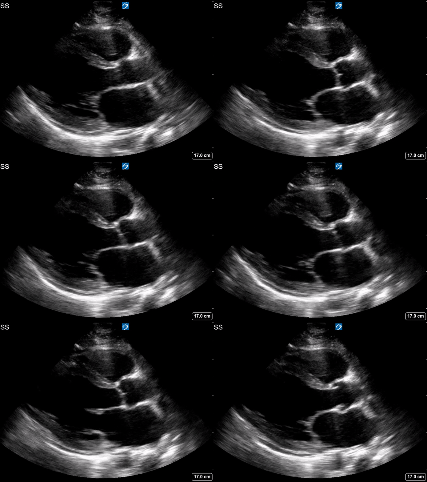
POCUS reveals the following:


Clips Gallery
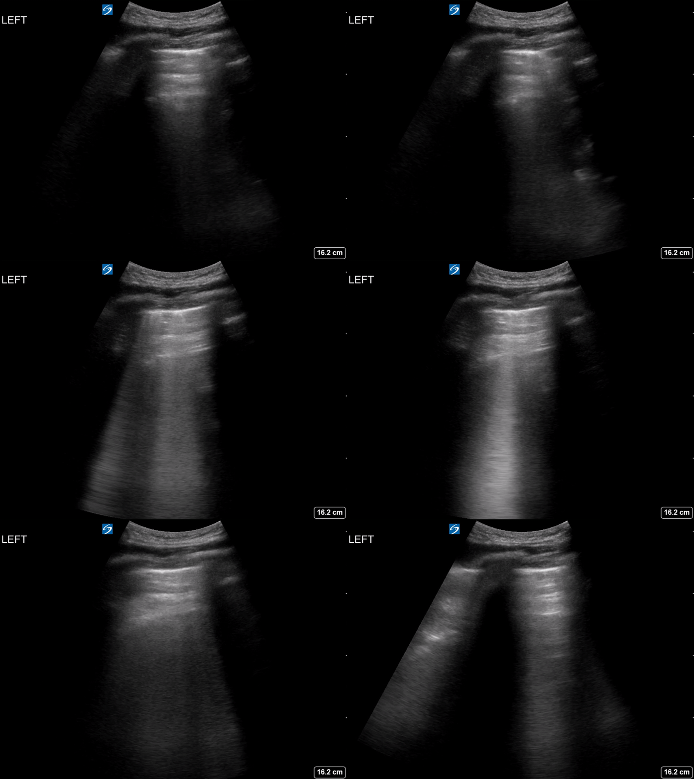
Clip 1- PLAX, Clip 2- PSAX, Clip3- A4C, Clip 4- Lung US, Left (L4)
-
EPSS (E-point septal separation) - >7mm suggests impaired LV function (see example below)
LV fractional shortening - can be “eyeballed” or measured in PLAX, should be approx 1/4 or 1/3
Assessing thickening/contraction of myocardial walls
MAPSE (mitral annular plane systolic excursion) - <8mm indicates impaired LV function
-
Severely impaired LV systolic function (estimated EF likely <15%)
-
TAPSE (tricuspid annular plane systolic excursion) - M-mode through tricuspid annular plane (free wall), <16mm indicates reduced RV longitudinal function (see example below)
RV size - normal RV size is approx 60-70% of LV. A 1:1 ratio definitely abnormal and may be acute or chronic. Obstructive causes of RV failure (eg PE, pHTN) often cause dilatation and bowing of the septal wall into the LV.
-
These are ring-down artefacts (also known as “B-lines” in the lung). They are formed by sound waves interacting with an air-fluid interface (gas bubbles).
Sound waves trapped within gas bubbles cause them to resonate and produce a secondary sound wave which returns to the transducer and produces a continuous non-attenuating hyperechoic laser-beam like artefact.
B-lines/ring-down artefact in the lung indicate increased lung density - often due to extravascular lung water but may also be caused by inflammation and fibrosis. When diffuse and bilateral, involving more than 2 lung zones bilaterally it is termed an alveolar-interstitial syndrome.
In this case, the B-lines were due to cardiogenic pulmonary oedema.
Case resolution
This patient had severe biventricular failure secondary to likely methamphetamine-related cardiomyopathy. He was admitted to CCU and had a departmental echocardiography, which confirmed the diagnosis.
Take-Home Message
Bedside focused echo (often complemented with lung ultrasound), even qualitative, is a good screening tool for a competent operator in selected cases as an extension of standard asessment.
Appendix
Image 1. EPSS of 23.9mm indicated severe LV impairment
Image 2. TAPSE of 11mm indicating reduced RV function


