
Case 20 - Torn Apart...
Author: Dr Nick Mani Reviewer: Dr Nish Cherian
A middle-aged man presents with a 1-week history of chest pain radiating to the back which came on suddenly during exertion and described as a ‘popping’ sensation.
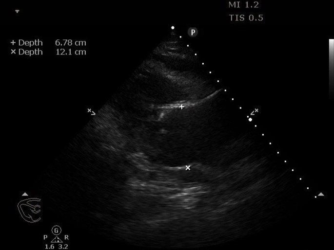
You elect to perform a bedside focused transthoracic echo with a dedicated suprasternal notch view as an extension of your standard clinical assessment:
Clip Collection (Press right/Left to change the clips)
Focused TTE PLAX, PSAX and SSN (other windows are limited)
TTE- Transthoracic Echocardiography, PLAX- Parasternal Long Axis, PSAX- Parasternal Short Axis, SSN- Suprasternal Notch
-
1- Thoracic Aortic Aneurysm
1- Pericardial Effusion (most likely haemopericardium in this case)
3- Bicuspid Valve
4- Intimal Flap (seen only in the the suprasternal notch view supported by doppler)
-
Presence of direct TTE signs (mainly intimal flap) had a sensitivity and specificity of 45.2% [95% confidence interval (CI) 37-53.6%] and 97.4% (95% CI 95.9-98.4%), while presence of any TTE sign, e.g. pericardial effusion or aortic valve regurgitation or aneurysm, had a sensitivity and specificity of 89% (95% CI 82.8-93.6%) and 74.5% (95% CI 71-77.7%).
Nazerian P, Mueller C, Vanni S, Soeiro AM, Leidel BA, Cerini G, Lupia E, Palazzo A, Grifoni S, Morello F. Integration of transthoracic focused cardiac ultrasound in the diagnostic algorithm for suspected acute aortic syndromes. Eur Heart J. 2019 Jun 21;40(24):1952-1960. doi: 10.1093/eurheartj/ehz207. PMID: 31226214.
-
Standard PSAX
and/or 2 chamber apical view
and/or proximal abdominal aorta view in longitudinal
-
Thoracic Aortic Syndrome/Dissection, (Type A) with background of undiagnosed thoracic aortic aneurysm and bicuspid valve, complicated by haemopericardium
-
CTA (unless haemodynamic instability unable to be optimised in which case TEE might need to be deployed)
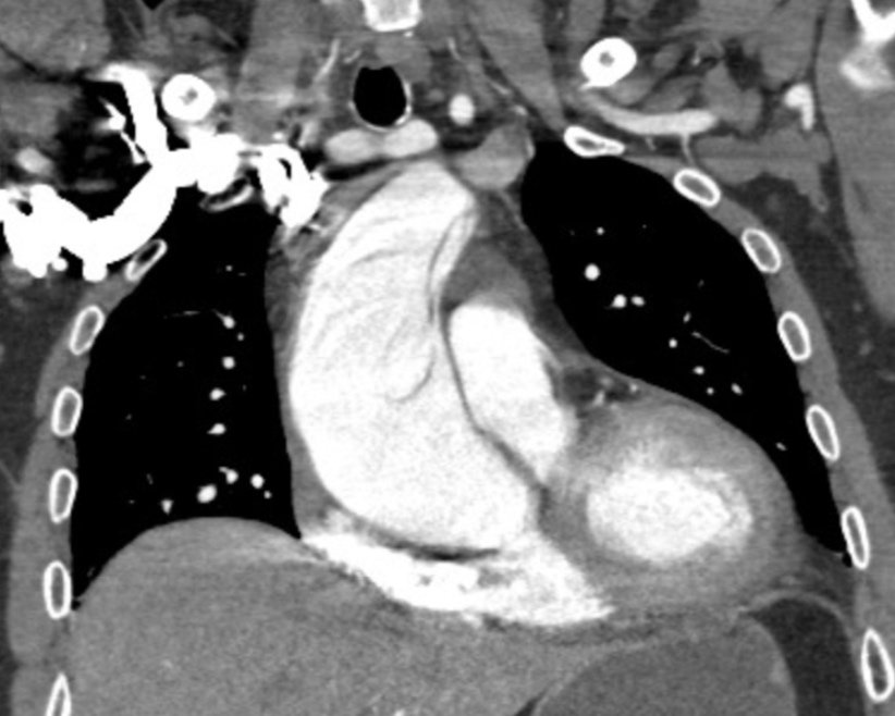
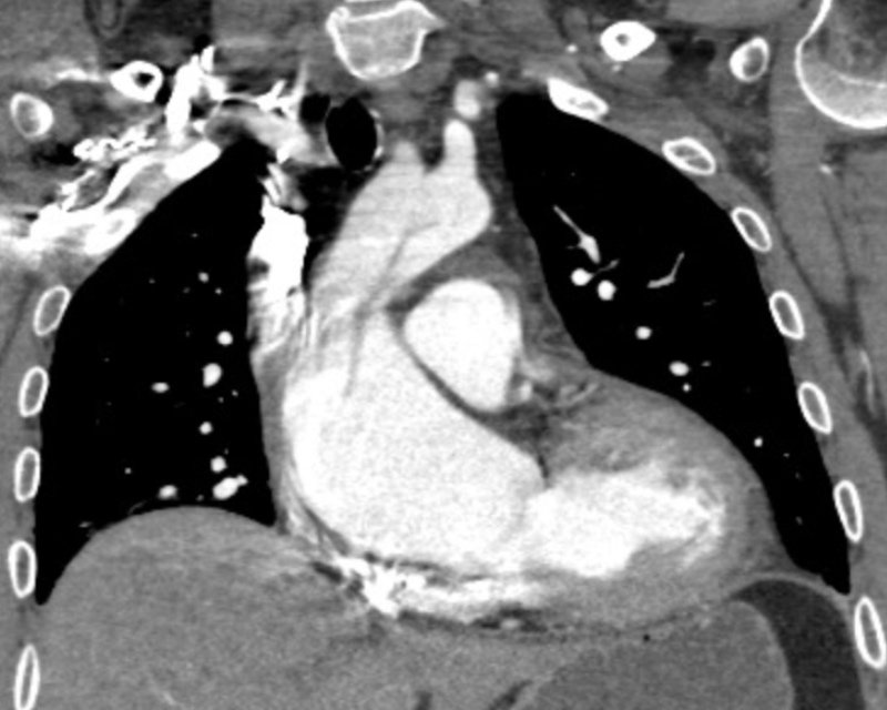
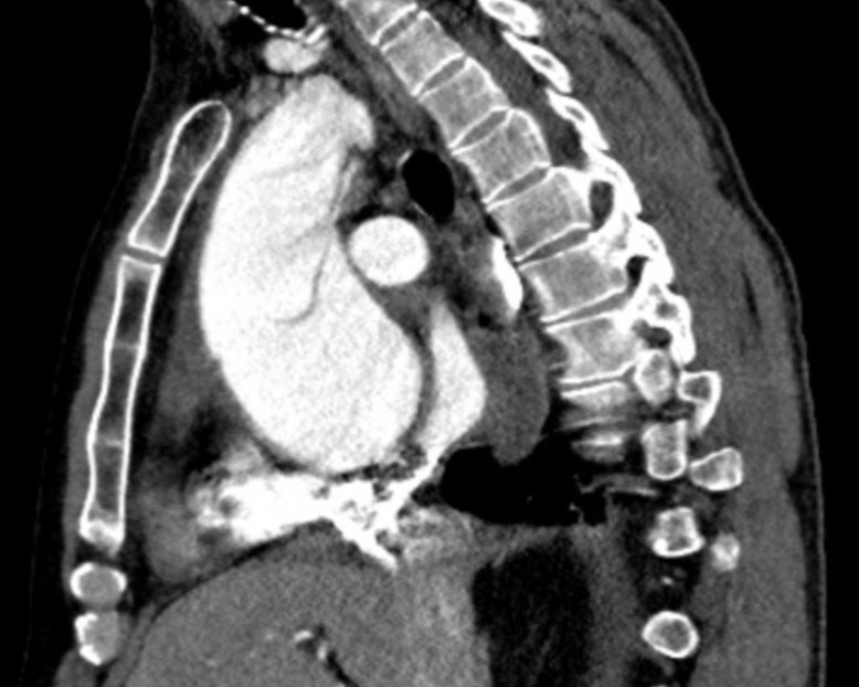
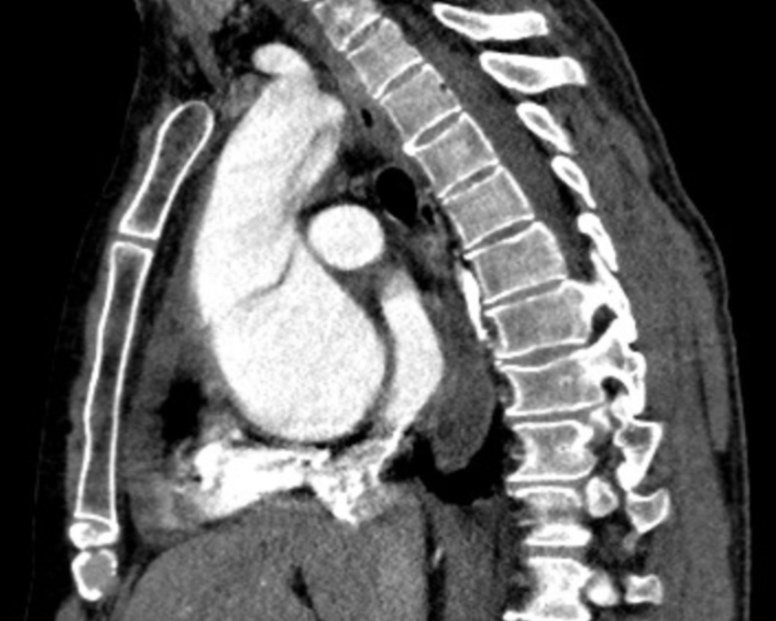
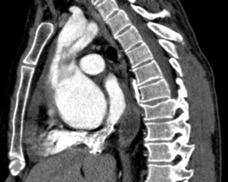
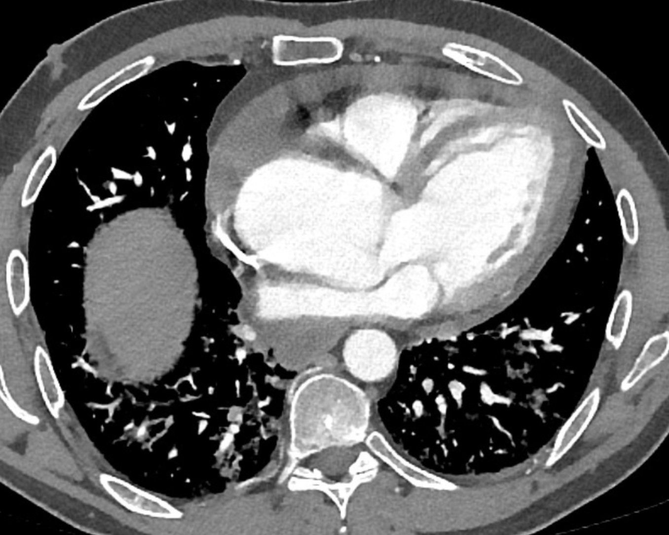


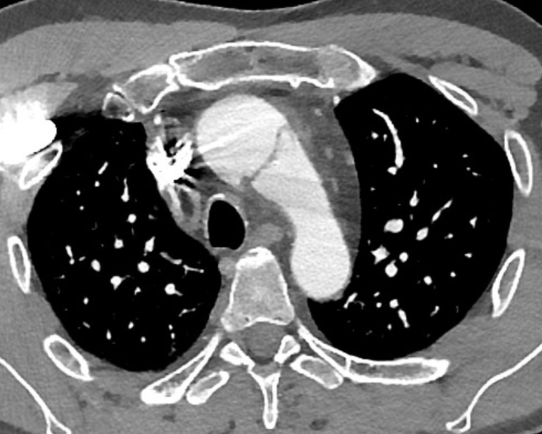
Clip Collection 2 (Press right/Left to change the clips)
CT Aortogram views confirms the diagnosis with early complication of haemopericardium
Case Resolution
The patient was urgently transferred to the regional cardiothoracic centre for surgical repair.
Take Home points
TTE could be useful as extension of clinical assessment particularly in haemodynamic instability, and/or resource limited settings
TTE should NOT be used to exclude and/or delay CT/transfer/surgery
CT Aortogram is the gold diagnostic standard