
Case 3 - Splash...
Author: Nish Cherian Reviewer: Nick Mani
A 70-year old male is urgently referred to ED by their GP with a stage 3 AKI incidentally noted on blood tests, as well as a pulsatile abdominal mass with concern for AAA.
The patient does not appear in much pain and has normal vitals in triage. On examination he does have a central abdominal mass extending above the umbilicus, which is slightly pulsatile.
POCUS reveals the following:

Image 1. Transverse view - transducer is in the central epigastric/umbilical region
-
Yellow arrow = abdominal aorta (lying above the vertebral body shadow and to the left of the IVC)
White arrow = a very enlarged bladder!

Image 2. Long axis view of the bladder
-
Grossly enlarged bladder with a diverticulum.
Clip 1. Left kidney
Clip 2. Right kidney
-
Severe bilateral hydronephrosis and hydroureter.
-
Hydronephrosis may be graded as mild, moderate or severe based on the degree of renal calyceal dilatation, blunting of renal pyramids and thinning of renal cortex.
-
There are a number of different formulas used to estimate bladder volume.
The prolate ellipsoid formula:
Volume = length x width x height x 0.52
Case resolution
The patient was catheterised in the ED and drained 2.5L of dark brown urine. He was admitted under Urology and had a departmental ultrasound later showing a large bladder haematoma (see below) causing a bladder outlet obstruction. CT did not reveal any malignancy or active haemorrhage. The patient initially had a cystoscopy and clot retrieval but continued to have haematuria and clot formation despite irrigation. This was thought to be platelet dysfunction secondary to uraemia. Bilateral urostomies were performed but despite this he became dialysis dependent for a few weeks. Following this his serum urea and haematuria improved and he was discharged home with a urinary catheter.
Appendices

Long axis view of bladder showing large intraluminal haematoma
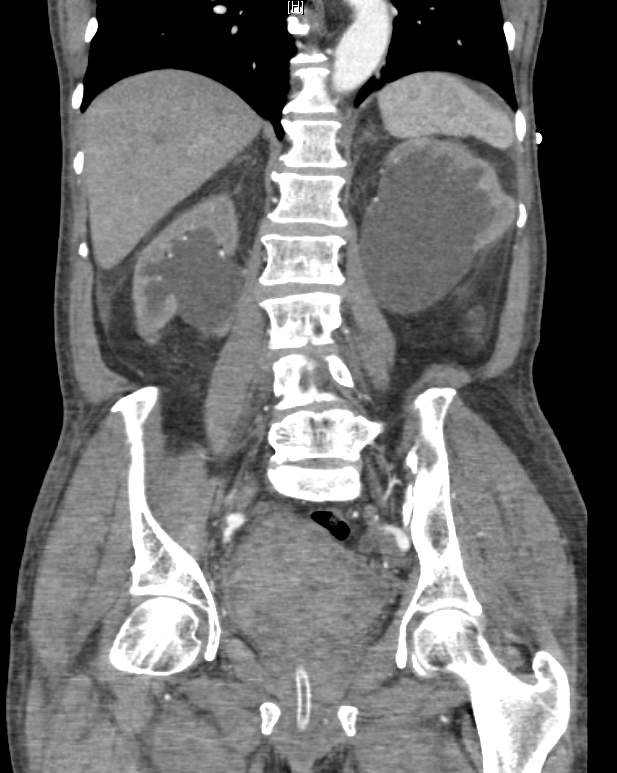
CT demonstrating severe bilateral hydronephrosis and hydroureter

POCUS 101 - Hydronephrosis grading