
Sound Bite 6 - Distal Radius Fracture (Paeds, 2 of 2)
Author: Dr Nick Mani
10yr old child presents to the Emergency Department with an injury to the left wrist after a fall. The wrist is swollen and tender with moderate dorsal deformity.
You elect to perform Point-of-Care Ultrasound (POCUS) of the right normal wrist and compare it to the left injured side: -

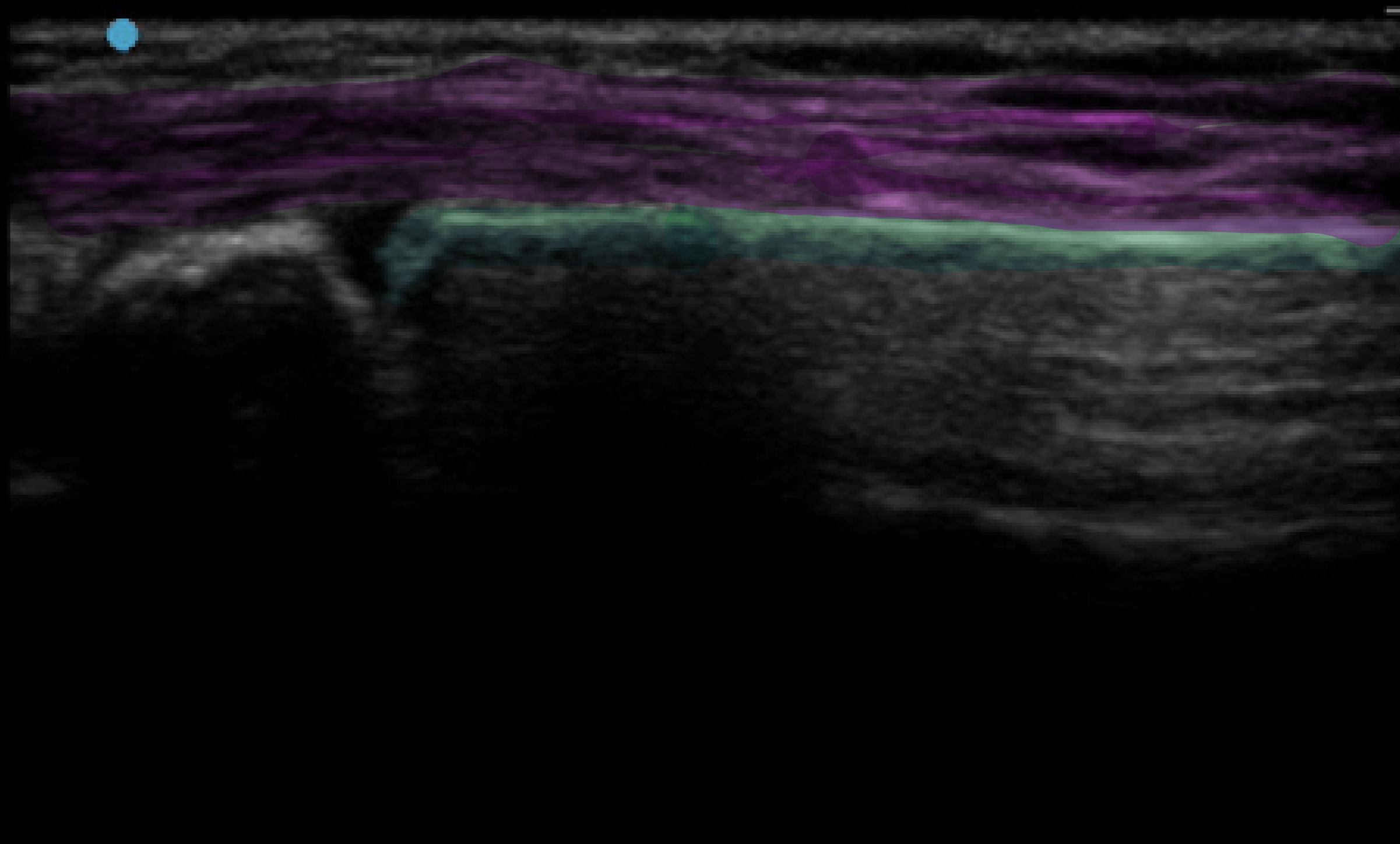
Normalogy- Right Distal Radius Dorsal Long Axis View
(Click the right arrow for annotation. Purple- Soft tissue, Green- Distal radial cortex. Note that gap on the left of the image is NOT a fracture, but the wrist joint)


Pathology- Left Distal Radius Dorsal Long Axis View of Cortical Fracture (Same Patient)
(Click the right arrow for annotation. Purple- Soft tissue, Green- Distal radial cortex, Orange- Distal radius cortex buckling)
Left distal radius palmer long axis view was also performed which demonstrated cortical fracture on the other side (not shown in here)
XR confirms the distal radius cortical fracture diagnosis, which is managed with closed manipulation/reducation and backstab: -
