Case 33 - Under Arrest…
Author: Dr Angus Perks Reviewer/Editor: Dr Nick Mani
A nurse from critical care bleeps you for an urgent review of a young patient on HDU due to a run of non-sustained VT. He has been on the unit for 3 days with COVID-19 pneumonitis, initially on high-flow FiO2 and now weaned down to 6L FiO2 via Hudson mask ready to be stepped down to a ward.
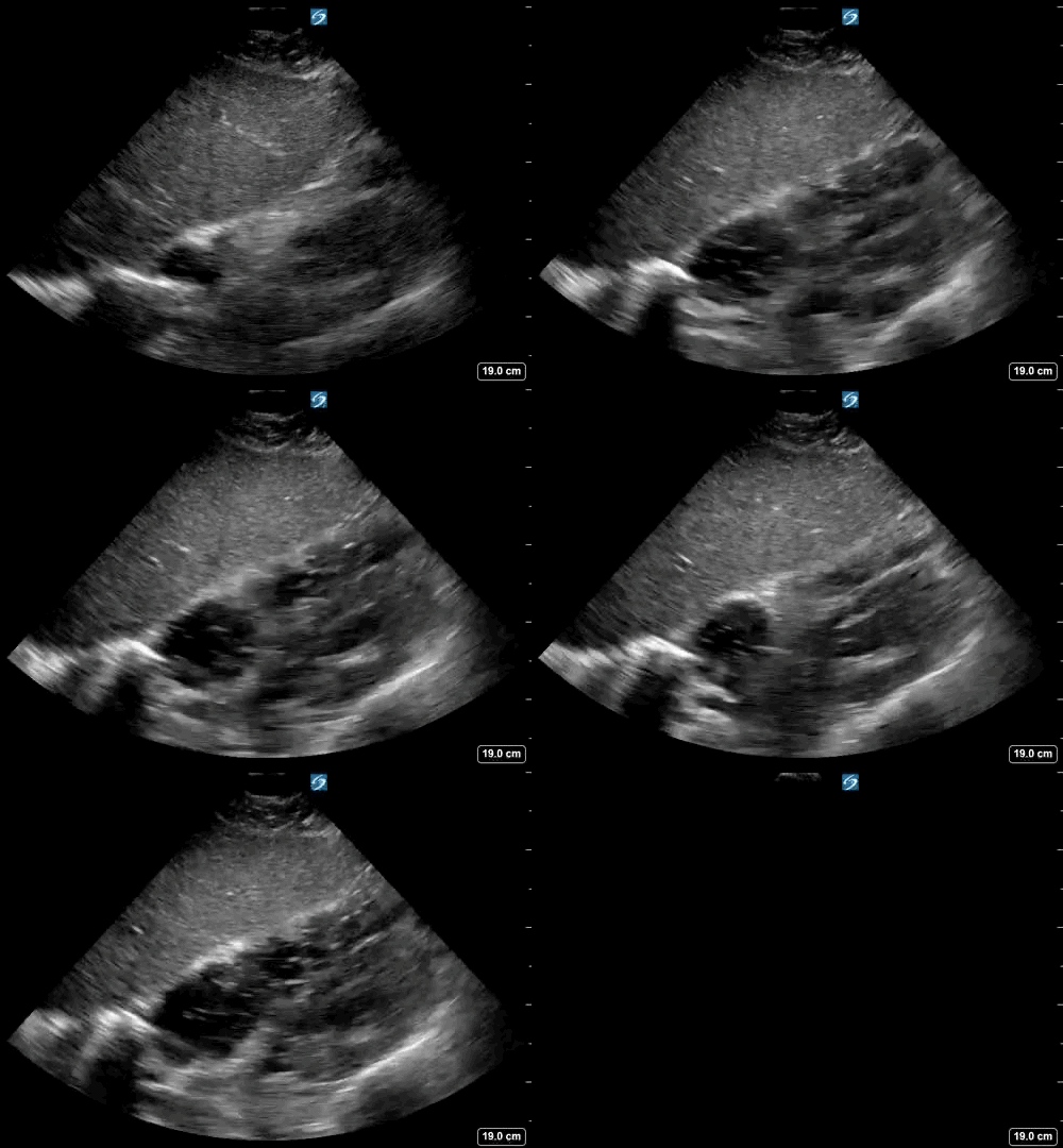
After you arrive at the unit and don PPE, the patient has just become unresponsive, ALS started with the 1st rhythm check showing pulseless electrical activity. You decide to be the dedicated performed of echo in life support (ELS). On the 2nd rhythm check the following echo loop is recorded:
Clip 1- 1st 10 sec recorded echo loop during the 2nd pulse check
-
This is a subcostal/subxiphoid 4 chamber view, one of the most commonly used views in cardiac arrest.
The advantage of this view is it allows the operator to position the probe even during compressions in preparation for a pause in CPR for a rhythm/pulse check.However, a 2021 study demonstrated superior images and shorter time to image acquisition using the parasternal long axis view. Of note this was with real patients but in simulated cardiac arrest - physiology and imaging may be different in true arrest!
-
Yes, the heart can be seen to beat.
-
Yes, rather than just slight ‘twitching’ of one area, there seems to be organised contratility, albeit slow and irregular.
-
Challenging question
Right at the beginning of the image, there is a suggestion the RV may be dilated, and at times the RA may appear to be pushing the inter-atrial septum into the LA. These features may suggest right heart failure, most likely given the history due to PE.
However, the RV is known to dilate in cardiac arrest of any etiology and so be wary of interpreting these signs as diagnostic certainty.
-
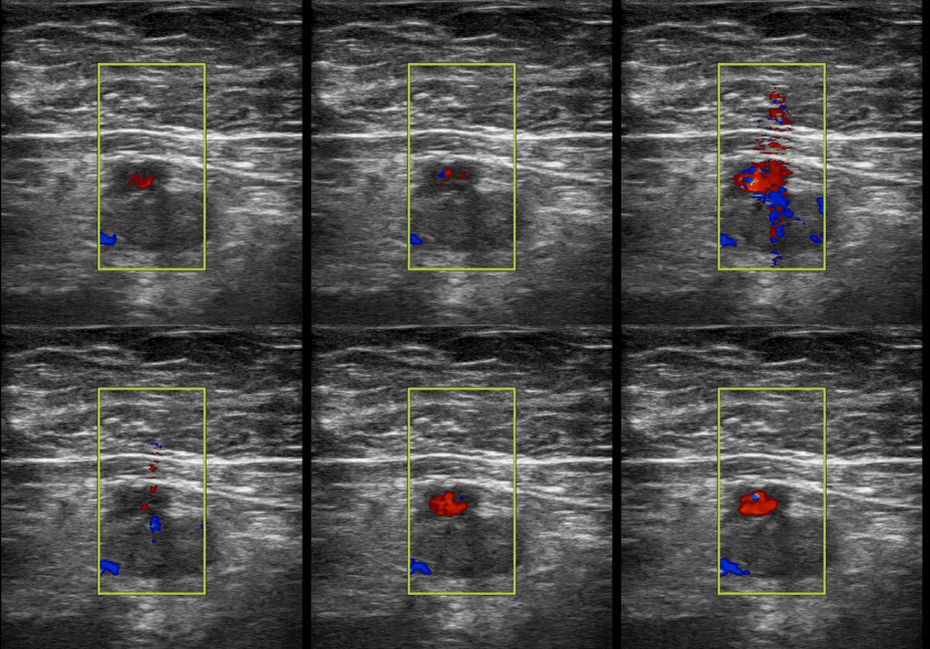
Yes. Common femoral vein ultrasound for DVT - see below.
Clip 1- Right common femoral vein POCUS
-
This is colour doppler over the common femoral vein (CFV) and artery (CFA).
Pulsatile flow is seen in the CFA but no flow is present in the CFV. Additionally there is hyperechoicity in the CFV suggestive of a thrombus.
-
This is a proximal DVT, which has a positive likelihood ratio of +13 for PE in a patient in this scenario.
Case Resolution
The patient received IV thrombolysis and a prolonged advanced life support of 90 minutes. No ROSC was achieved at any point and resuscitation was stopped.
Take-home Message
Echo in life support could help identify fine VF and pseudo-PEA. It could guide the correct depth and placement of chest compressions to avoid inadvertent left ventricular outflow tract obstruction by misplaced compression.
In non-shockable rhythm, it could help to identify reversible causes such as hypovolaemia, PE, and pericardial effusion/tamponade. Additional POCUS scan might be also is required to rule in AAA, free fluid in the chest/abdomen/pelvis, and proximal DVT.


