
Case 32- Octopus Trap
Author: Dr Salman Naeem Reviewer: Dr Suneeta Teckchandani/Dr Nick Mani
A 69 year old female presents to the emergency department with 5 days of worsening shortness of breath with orthopnoea. This seems to have coincide by her husband recent severe stroke.
Vital signs are 98% OA, RR26, 97bpm, 128/76. There are some signs of congestive cardiac failure on examination. There was non-specific signs of ischemia throughout ECG leads, CXR and bloods including troponin awaiting.
You decide to perform a bedside focused echo as an extension your clinical assessment: -
Clip collection - Bedside Focused Echo (click right/left arrows to cycle between clips)
-
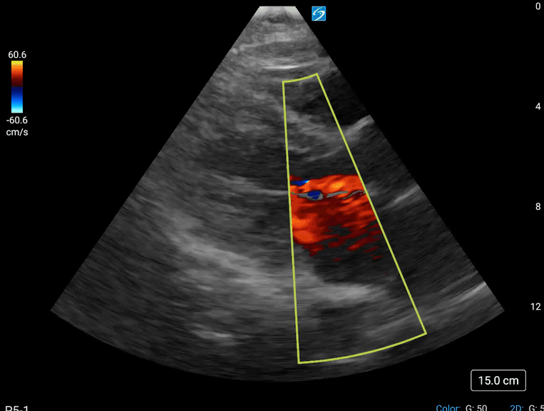
1st clip- Parasternal Long Axis
2nd clip- Parasternal short axis
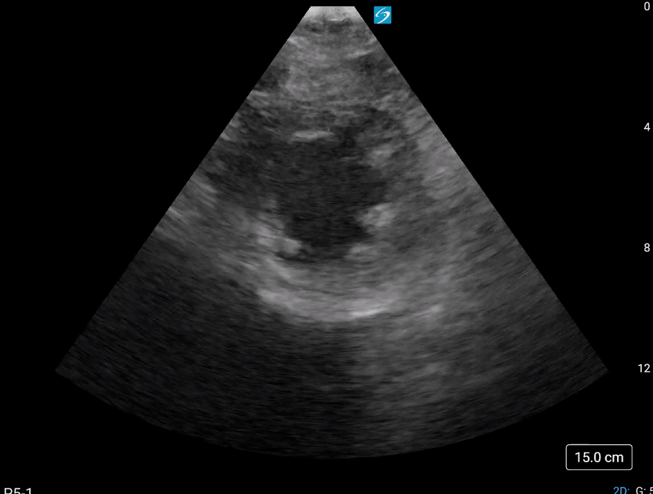
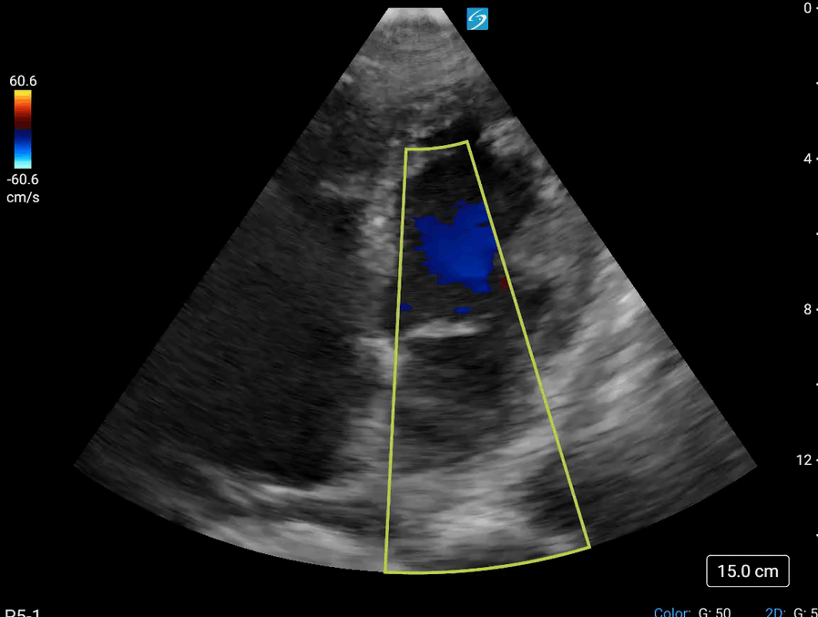
3rd clip- Apical 4 chambers
-
1st clip demonstrates akinesia (wall thickening of <10%) of the anteroseptal and inferolateral (posterior) wall with reduced excursion of the mitral valve leaflets and jet of mitral regurgitation. Note the colour doppler window is spanning through Aortic and mitral valve to provide an overview. Ideally separate window for each valve is required. Note the pleural effusion present left side.
2nd clip shows all the walls of LV are akinetic at papillary muscle level.
3rd clip illustrates akinesia of the inferior septal wall and lateral wall of the LV with mitral regurgitation. Notice that the apex is not viewed well on this window as it is foreshortened. The RWMA (regional wall motion abnormality) is sparing of very small sections of the basal segments.
-
Considering the acute history related to sudden stress in life, the diagnosis is likely to be Takotsubo/stress cardiomyopathy though acute coronary syndrome needs to be excluded first.
CASE RESOLUTION
CXR demonstrated signs of CCF including pleural effusion left side, and serial troponin tests were normal. The patient was admitted to CCU where she had delayed PCI and Left ventriculography, which confirmed the final Takotsubo or stress cardiomyopathy.
Take Home Message
Takotsubo or stress cardiomyopathy is not uncommon, and has a distinct appearance on echo. However Coronary artery disease should be excluded as a cause before such before such diagnosis is made.
Other notes
Takotsubo or stress cardiomyopathy is characterised by a sudden onset of severe and transient LV dysfunction due to catehcholamine surge secondary to stress, typically seen in post-menopausal women. Systematic evaluation by two- dimensional echocardiography reveals a distinctive pattern of LV contractility in Takotsubo cardiomyopathy characterized by symmetrical RWMA extending equally into the anterior, inferior, and lateral walls and not confined to a particular coronary artery territory (1). The usual echo picture is hypo/a kinesia of the apex of the LV with normal basal LV contraction with a typical picture of apical ballooning. However, in reverse Takotsubo patient has hypokinesia of the basal LV with normal contractility of the apex.
The name "takotsubo" comes from the Japanese word takotsubo "octopus trap" as the left ventricle of the heart takes on a shape resembling an the trap when affected by this condition.
References
1. Rodolfo Citro, Fausto Rigo, Quirino Ciampi, Antonello D’Andrea, Gennaro Provenza, Marco Mirra, Roberta Giudice, Francesco Silvestri, Giuseppe Di Benedetto, and Eduardo Bossone. Echocardiographic assessment of regional left ventricular wall motion abnormalities in patients with tako-tsubo cardiomyopathy: comparison with anterior myocardial infarction. European Journal of Echocardiography (2011) 12, 542–549 doi:10.1093