Case 30 - The Mysterious Lump…
Author: Dr Salman Naeem Reviewer: Nish Cherian/Nick Mani
A 44-year old male with no past medical history presents to the Emergency Department with sudden onset abdominal pain in the umbilical region, associated with vomiting which started 4 hours ago.
On arrival HR 110/min. He is in severe pain despite 10mg of IV morphine. The triage nurse asks for a clinical review.
On examination he has a small swelling protruding from the umbilicus which is very tender and not reducible. The rest of the abdomen is soft and non-tender.
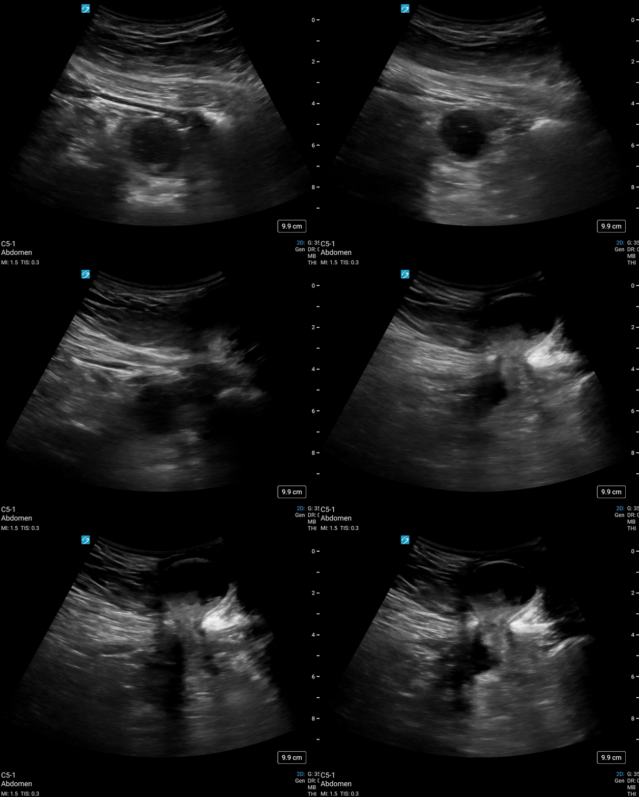
A bedside POCUS examination is performed of the swelling with the clips as seen below:-


-
Scan of the umbilical hernial sac showing herniation of small bowel into the umbilical hernia. As one follows the small bowel one can see that there is protrusion of the small bowel into the sac.
Scanning the bowel around the umbilicus shows early dilatation of small bowel (b) in Image 2. This would support incarceration of the hernia.
-
Image 1 shows rectus muscle of the abdominal wall (a), defect in the abdominal wall (b) and hernial sac containing bowel contents (c).
Image 2 shows rectus muscle (a), small bowel (b) and subcutaneous tissue (c)
-
The most common hernias are due to defect in the abdominal wall. Using linear probe scan the hernial sac and then trace the origin of sac with the ultrasound. It is important to scan for and document contents of the sac, reducibility and size of the defect in the abdominal wall. Sometimes hernias are not visible in supine position, and one might have to stand the patient up and ask them to increase intra-abdominal pressure by asking them to cough to visualise the hernial sac and its contents.
If the defect in the abdominal wall is small then it is more likely that the hernial contents will not be reduced spontaneously and patient will need surgical decompression.
For more information on scanning for hernias follow this link https://www.ncbi.nlm.nih.gov/pmc/articles/PMC9262670/
CASE RESOLUTION
The POCUS findings were shown to the Surgical Registrar who bypassed CT and took the patient straight to theatre. Intra-operatively, a strangulated hernia was confirmed, but the small bowel was still viable. The hernia was decompressed and the abdominal wall defect closed. The patient was discharged home the following day.
TAKE HOME MESSGAE
Strangulated hernia is a surgical emergency and POCUS enhanced examination can be quite sensitive in diagnosing hernia, its contents and associated ileus, reducing exposure to ionizing radiation and expediting definitive care.
References:-
Wu WT, Chang KV, Lin CP, Yeh CC, Özçakar L. Ultrasound imaging for inguinal hernia: a pictorial review. Ultrasonography. 2022 Jul;41(3):610-623. doi: 10.14366/usg.21192. Epub 2022 Feb 24. PMID: 35569836; PMCID: PMC9262670.
Tamburrini S, Serra N, Lugarà M, Mercogliano G, Liguori C, Toro G, Somma F, Mandato Y, Guerra MV, Sarti G, Carbone R, Tammaro P, Ferraro A, Abete R, Marano I. Ultrasound Signs in the Diagnosis and Staging of Small Bowel Obstruction. Diagnostics (Basel). 2020 May 3;10(5):277. doi: 10.3390/diagnostics10050277. PMID: 32375244; PMCID: PMC7277998.
Lin, Y. C., Yu, Y. C., Huang, Y. T., Wu, Y. Y., Wang, T. C., Huang, W. C., Yang, M. D., & Hsu, Y. P. (2021). Diagnostic accuracy of ultrasound for small bowel obstruction: A systematic review and meta-analysis. European journal of radiology, 136, 109565. https://doi.org/10.1016/j.ejrad.2021.109565
